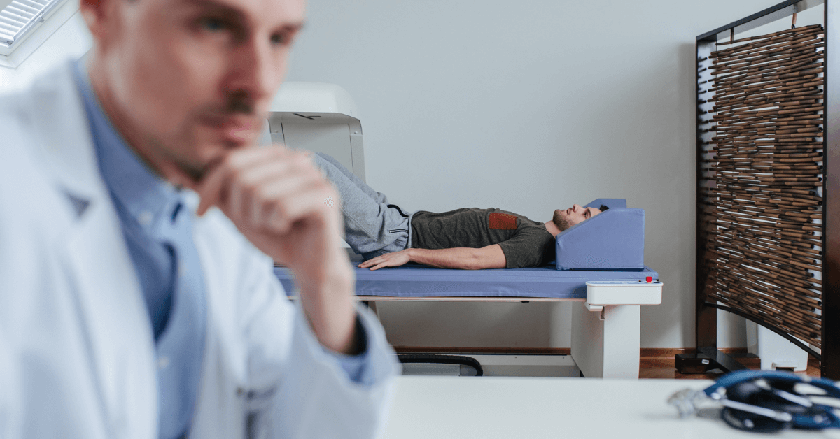
A DXA (Dual-Energy X-ray Absorptiometry) scan is a medical imaging technique primarily used to assess bone mineral density (BMD), helping to diagnose conditions like osteoporosis and to evaluate fracture risks. While it’s a common and non-invasive procedure, one question that often arises is whether it’s safe due to the radiation involved. Given that radiation exposure is a significant concern in medical imaging, it’s important to understand how the DXA scan works and whether its radiation exposure poses a risk to health.
In this blog, we’ll explore how the DXA scan works, the amount of radiation involved, and whether it’s considered safe for patients.
How Does the DXA Scan Work?
A DXA scan uses two low-dose X-rays at different energy levels to pass through the bones and soft tissue of your body. The machine compares the amount of X-ray absorbed by the bones versus the soft tissue, which provides precise measurements of bone mineral density (BMD). This measurement helps determine the strength and health of your bones and is a key tool in diagnosing conditions like osteoporosis or assessing the risk of fractures.
Typically, the scan focuses on the spine, hip, and sometimes the forearm. The procedure is painless, quick, and typically takes only about 10-30 minutes, depending on the area being scanned.
Radiation in the DXA Scan: How Much Is There?
One of the major concerns with any medical imaging procedure is the amount of radiation involved. However, DXA scans are among the least invasive and lowest radiation techniques used in diagnostic medicine.
Radiation dose comparison: A typical DXA scan uses approximately 1/10th of the radiation of a standard chest X-ray. To put this into perspective, the radiation dose from a DXA scan is very small—typically around 1 to 5 microsieverts (µSv). For comparison, a chest X-ray delivers around 100 µSv, and the average person is naturally exposed to about 2,000 µSv of background radiation per year just from environmental sources like the sun, soil, and cosmic rays.
Radiation in context: To provide a clearer understanding of the exposure, the amount of radiation you’d receive during a DXA scan is roughly equivalent to the radiation exposure you get from a few days to a week of natural background radiation. As a result, it is considered to be a very low-risk procedure in terms of radiation exposure.

Is the DXA Scan Safe?
Given the low radiation dose involved, DXA scans are generally considered very safe. However, as with any medical procedure involving radiation, there are some factors to consider.
- Risk vs. Benefit
For most patients, the benefits of a DXA scan far outweigh the risks. If you are at risk for osteoporosis or have a family history of bone-related issues, the ability to accurately assess your bone density could significantly improve your healthcare management and preventive care. Diagnosing conditions like osteoporosis early can help prevent fractures and other complications associated with weakened bones.
For women over the age of 65, or postmenopausal women under 65 with risk factors, a DXA scan is often recommended to detect bone loss before it leads to fractures. Similarly, men over 70 and individuals with certain medical conditions, such as arthritis, diabetes, or chronic kidney disease, might also be candidates for this scan.
The dose of radiation from a DXA scan is so low that the potential risk of developing cancer from this exposure is considered negligible. In fact, a study by the Radiation Protection of Patients (IRPA) organization concluded that the cancer risk from a DXA scan is far smaller than the potential benefits of diagnosing and treating bone health conditions early.
While DXA scans are considered safe for most individuals, there are some groups for whom the procedure may not be necessary or recommended:
Pregnant women: DXA scans use radiation, so pregnant women should generally avoid the procedure, unless it’s deemed absolutely necessary by a healthcare provider. If you are pregnant or suspect you may be, it’s important to inform the technician or doctor before scheduling the scan.
Young children: While DXA scans are safe for children when medically necessary, they are typically not recommended unless there is a significant reason to assess bone health. This is because children are more sensitive to radiation, and the potential risks may outweigh the benefits unless there’s a pressing medical reason.
- Minimizing Radiation Exposure
While DXA scans already involve minimal radiation, healthcare providers typically take extra precautions to ensure the lowest possible exposure. For example, the technician may use protective shields or minimize the number of areas being scanned to limit the dose even further.
Additionally, because DXA scans are typically used for ongoing monitoring of bone density in patients at risk, it’s important that healthcare providers only recommend them when appropriate and spaced out at reasonable intervals, rather than conducting them too frequently.
The Role of DXA Scans in Bone Health
While radiation is a concern with any medical imaging procedure, the low dose of radiation used in DXA scans makes them a safe and effective tool for diagnosing and monitoring bone health. In addition to helping detect conditions like osteoporosis, DXA scans can also be useful in:
Assessing fracture risk: By measuring bone density, a DXA scan helps predict the likelihood of bone fractures, which is especially important for elderly individuals or those with conditions that affect bone strength.
Monitoring bone loss: For patients with osteoporosis or other bone diseases, regular DXA scans can track the progression of bone loss and help guide treatment plans, ensuring that patients receive appropriate interventions to reduce fracture risk.
Evaluating treatment effectiveness: DXA scans are also used to evaluate how well osteoporosis treatments are working, allowing doctors to adjust treatment plans as needed.
Conclusion
The DXA scan is an important diagnostic tool for assessing bone mineral density, especially for individuals at risk of osteoporosis or fractures. While it does involve a small amount of radiation, the dose is so low that the procedure is considered very safe for most patients. The benefits of early detection and monitoring of bone health far outweigh the risks associated with the minimal radiation exposure involved. As always, if you have concerns about radiation or your health, it’s a good idea to discuss them with your healthcare provider, who can help you weigh the risks and benefits based on your specific health needs.
Schedule Your Appointment Today
Schedule your DXA scan at DXA Body Composition NC. And take the first proactive step towards understanding and optimizing your health. Our process is quick, painless, and performed on an outpatient basis. The machines we use are comfortable and open, ensuring a stress-free experience.
Don’t let your health journey be a mystery; let DXA Body Composition NC be your guide. Take the next step by scheduling your DXA scan appointment. Contact us today to discover the precision, accuracy, and personalized insights that will propel you toward your health and fitness goals. Your journey to optimal well-being starts here.
