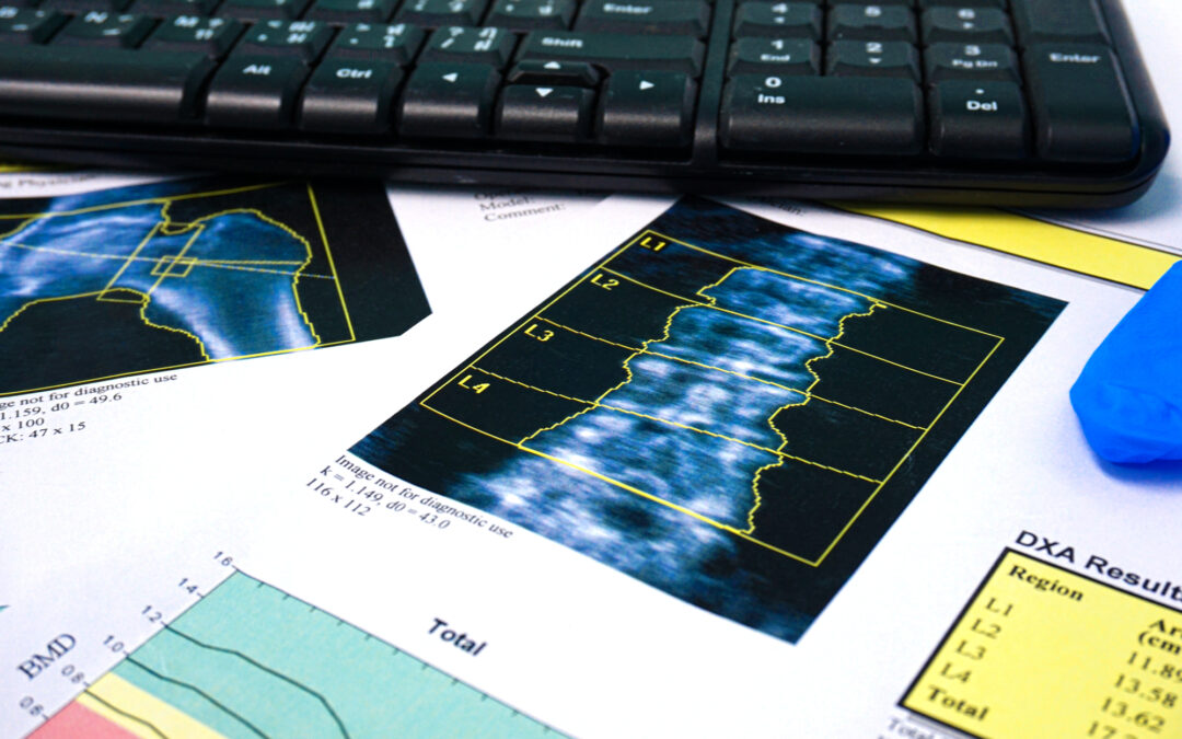
The radiation dosage administered in DXA scans is minimal, comparable to the levels employed in routine X-rays. While daily exposure to ionizing radiation occurs naturally in our environment, additional exposures can marginally elevate the risk of cancer development later in life.
Utilizing dual X-ray absorptiometry (DXA), bone density – encompassing both thickness and strength – is quantified by directing high and low-energy X-ray beams (a variant of ionizing radiation) through the body, typically targeting the hip and spine regions. This diagnostic procedure holds significance in identifying osteoporosis or bone thinning, with the potential for repetition over time to monitor fluctuations in bone density.
A DXA scan falls within the realm of medical imaging tests, employing minimal levels of X-rays to assess bone density. DXA, an acronym for “dual-energy X-ray absorptiometry,” is widely acknowledged by medical professionals as the most practical, straightforward, and cost-effective diagnostic tool for osteoporosis.
What is the typical radiation dosage for a (DXA) bone scan?
These examinations are widespread, and inquiries regarding the associated X-ray doses are often deflected due to their extremely low levels. While it’s accurate that these scans, technically referred to as DXA, yield some of the lowest effective doses among X-ray procedures in medicine, there is no reason not to explicitly disclose these figures.
The primary reason for the remarkably low dose in DXA scans compared to other skeletal X-rays is the deliberate use of minimal radiation, sufficient only for detecting variations in the transmission of two different energy levels of X-rays through bones.
Several methods exist for quantifying radiation dose, with the Sievert (Sv) being the most widely employed unit. The Sievert considers both the energy deposited by radiation and the specific body part exposed, providing a measure that reflects the overall potential harm to cells in the body.
In the context of radiation therapy, where the goal is to target and harm tumor cells, doses in the order of several Sv are utilized. In contrast, diagnostic radiology aims for the smallest doses essential to generate precise X-ray images of the body, requiring significantly less radiation, typically measured in thousandths of sieverts or millisieverts (mSv).
Below are the effective doses linked to DXA scan radiation and several other diagnostic radiology exams for reference:
- DXA—Whole body: 0.003 mSv
- Dental X-ray: 0.005 mSv
- Chest X-ray: 0.1 mSv
- Abdomen and Pelvis CT: 10 mSv
What constitutes the permissible threshold for radiation scans within a year?
If you are having multiple medical examinations, the accepted limit is 20 mSv per year. This is measured in “mSv,” denoting 1000 millisieverts — a unit that, while not as extensive as a standard Sievert, is significantly higher than the typical range of 1-4 uSv for a single DXA scan.
What are the benefits vs. risks?
It is important to weigh the potential risks of not having a DXA scan. Overlooking visceral fat percentages might result in metabolic syndrome. A delayed response to sarcopenia, characterized by the juxtaposition of muscle loss and excess weight in aging individuals, can impede cognitive function.
Considering the trade-off between the risk of significant physical ailments and the benefits of enhanced health and wellness, the equivalent of one-tenth of the radiation from a cross-country flight seems like an acceptable trade-off.
What To Expect At Your DXA Appointment
Preparation for a DXA scan is straightforward and generally requires no special measures. However, it is crucial to inform both your doctor and the technologist if there is a chance of pregnancy or if you have recently undergone a barium exam or received contrast material for a CT or radioisotope scan.

Throughout the procedure, you might be requested to remove jewelry, eyeglasses, and any attire that could disrupt the imaging process.
Lite, thin material should be worn for the scan. For men, shorts and a light T-shirt are acceptable. For women, yoga-style clothing tends to be best. Do not wear jeans, wool, or heavy material for your scan.
You will recline on a table, maintaining stillness as the images are captured. Typically lasting around 15-20 minutes, the procedure concludes with your healthcare provider providing follow-up information on your test results and, if required, initiating a treatment plan.
It’s also recommended to abstain from taking calcium supplements for at least 24 hours before the scheduled exam.
Schedule Your DXA Scan with DXA Body Composition NC
Our body scans can assist patients who want to enhance or monitor their fitness, assess their bone health, or just want to understand their body composition.
Seek guidance from a healthcare provider to discuss your concerns and any other health-related questions you may have. Take your first proactive step of finding a provider and scheduling an appointment for a potential improvement in your health.
For more information about our DXA machines and DXA scan radiation, reach out to DXA Body Composition NC today!
